暗闇の中で視界を確保する「ナイトビジョン」は、生物学的な適応と高度な工学的アプローチの組み合わせによって実現しています。人間にとっての暗視とは、極めて少ない光を効率的に捉える能力、あるいは可視光以外の電磁波を検知して視覚化することを指します。
Key Facts
- 生物学的暗視:桿体細胞(かんたいさいぼう)による低光量検知と、一部の動物が持つ反射層「タペタム」が鍵となる。
- 画像増幅:微弱な光子を電子に変換し、増幅させて可視化する技術。
- 能動的照射:赤外線などの不可視光を照射し、それをセンサーで捉える手法。
- 熱線映像:物体が放出する熱(赤外線)の温度差を画像化する技術。
暗視を決定づける2つの要素
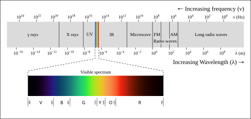
暗視能力は、主に「スペクトル範囲」と「強度範囲」という2つの側面から定義されます。スペクトル範囲とは、どの波長の光を感知できるかということであり、人間は電磁スペクトルのごく一部である可視光しか見ることができません。一方、強度範囲とは、極めて微量な光であっても視認できる能力を指します。
生物による視覚の拡張
多くの動物は人間よりも優れた暗視能力を持っています。これは、眼球やレンズのサイズが大きいことや、網膜に存在する光受容細胞のうち、低光量に反応する桿体細胞の割合が非常に高いことなどが理由です。また、猫や犬などの夜行性動物は、網膜の背後に「タペタム(輝膜)」という反射組織を持っており、一度通り過ぎた光を再び網膜に反射させることで、光の利用効率を最大化しています。これが、夜間に動物の目が光って見える「アイシャイン」の正体です。
![Normalised absorption spectra of the three human photopsins and of human rhodopsin (dashed). Drawn after Bowmaker and Dartnall (1980).[5]](/images/93/b1/93b18a2e590fd678858acceac4c1aea461976c738ed342c352fb111c2d7a825e.webp)
人間の目の仕組みと暗所への適応
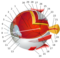
人間の網膜には、色を識別する錐体細胞と、暗い場所で機能する桿体細胞があります。暗所では桿体細胞に含まれるロドプシンというタンパク質が重要な役割を果たします。ロドプシンは光を吸収すると形状が変化し、視覚信号を誘発しますが、一度消費されると肝臓で再生され、再び目に供給されるまで時間がかかります。
完全に暗闇に適応するには約45分かかりますが、大部分の適応は最初の5分間で完了します。また、ロドプシンは長い波長の赤色光に反応しにくいため、夜間の操縦や作戦時に赤色の照明が使われます。これにより、暗所視能力を維持したまま計器類を確認することが可能です。
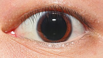
瞳孔の散大も光の取り込み量を増やす生物学的プロセスの一つです。通常、明るい場所では2mm程度まで収縮しますが、暗所では最大8〜9mmまで拡大し、より多くの光を網膜に届けようとします。
現代の暗視テクノロジー
テクノロジーによる暗視は、大きく分けて「画像増幅」「能動的照射」「熱線映像」の3つの方式に分類されます。
1. 画像増幅(イメージ・インテンシファイア)
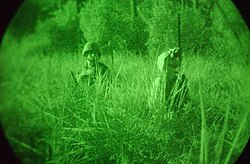
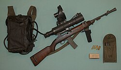
星明かりや月光などの極めて微弱な自然光を増幅させる方式です。真空管を用いた画像増幅管(フォトマルチプライヤー)が使用され、光子が光電陰極に当たると電子が放出され、それがマイクロチャネルプレートで増幅され、最終的に蛍光スクリーンに衝突して可視光として表示されます。
2. 能動的照射(アクティブ・イルミネーション)
人間には見えない近赤外線(NIR)や短波赤外線(SWIR)を照射し、それを専用のセンサーやCCDカメラで捉える方式です。高解像度な画像が得られるため、住宅や政府施設のセキュリティカメラに広く採用されています。ただし、赤外線センサーを持つ相手には照射光が見えてしまうため、軍事的な隠密作戦には不向きな側面があります。
3. 熱線映像(サーマル・ビジョン)
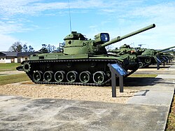
光ではなく、物体が放出する熱(赤外線)の温度差を検知する方式です。光源が全くない完全な暗闇でも機能し、霧や煙をある程度透過して視認できるため、航空機の前方監視赤外線(FLIR)などに利用されています。ただし、ガラスやアクリルなどの素材は赤外線を遮断するため、壁や窓越しに内部を見ることはできません。
暗視装置の進化と現状
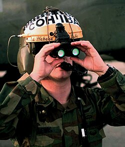
かつての暗視手段は、大きなレンズで光を集める「ナイトグラス(夜間用眼鏡)」が主流でしたが、重量とサイズが課題でした。現代では、軍用としてPVS-14のようなモノキュラー(単眼式)や、特殊部隊向けのGPNVG-18のようなクアッドチューブ(四眼式)などが運用されています。
また、最新のデジタル暗視では、高感度なsCMOS(科学用CMOS)センサーが採用されており、人間の目の感度を遥かに超える視認性を実現しています。これらの技術は軍事のみならず、航空機の状況認識システム(EVS)や高級車の運転支援システムとしても応用されています。
| 方式 | 光源 | 原理 | 主な特徴 |
|---|---|---|---|
| 画像増幅 | 自然光(月・星) | 光子を電子に変換し増幅 | 受動的で隠密性が高い |
| 能動的照射 | 赤外線照射器 | 不可視光の反射を検知 | 高解像度だが位置が露呈しやすい |
| 熱線映像 | 物体自体の熱 | 温度差を画像化 | 完全な暗闇や煙の中でも視認可能 |
Frequently Asked Questions
なぜ夜間に赤いライトが使われるのですか?
網膜の桿体細胞にあるロドプシンという物質が、長い波長の赤色光に反応しにくいためです。赤色光を使用することで、暗闇に適応した状態(暗所視)を維持しながら視界を確保できます。
サーマルカメラで壁の向こう側は見えますか?
いいえ、見えません。サーマルカメラは物体の表面温度を検知するものであり、壁やガラスなどの固体は赤外線を透過させないため、遮蔽物の向こう側を透視することは不可能です。
動物の目が夜に光るのはなぜですか?
網膜の背後に「タペタム(輝膜)」という反射層を持っているためです。光を反射させて網膜をもう一度通過させることで、少ない光でも効率的に捉えることができます。
デジタル暗視とアナログ(増幅管)暗視の違いは何ですか?
アナログ方式は真空管で光を物理的に増幅させますが、デジタル方式は高感度CMOSセンサーで光を電気信号として捉え、デジタル処理して表示します。デジタル方式は記録や他のシステムとの連携が容易という利点があります。
人間が暗闇に完全に慣れるまでどのくらい時間がかかりますか?
ロドプシンの再充填に時間がかかるため、完全に適応するには約45分ほどかかります。ただし、視覚的な感度の大部分は最初の5分程度で向上します。
References
- Chijiiwa, Taeko; Ishibashi, Tatsuro; Inomata, Hajime (1990). "Histological study of choroidal melanocytes in animals with tapetum lucidum cellulosum (abstract)". Graefe's Archive for Clinical and Experimental Ophthalmology. 228 (2): 161–168. :10.1007/BF00935727. 2338254. 11974069.
- Milius, Susan (2012). "Mantis shrimp flub color vision test". . 182 (6): 11. :10.1002/scin.5591820609. 23351000.
- "The Human Eye and Single Photons".
- "Sensory Reception: Human Vision: Structure and function of the Human Eye" vol. 27, p. 179 Encyclopædia Britannica, 1987
- Bowmaker, J K; Dartnall, H J (1 January 1980). "Visual pigments of rods and cones in a human retina". The Journal of Physiology. 298 (1): 501–511. :10.1113/jphysiol.1980.sp013097. 1279132. 7359434.
- Luria, S.M.; Kobus, D.A. (April 1985). "Immediate Visibility after Red and White Adaptation" (PDF). Submarine Base, Groton, CT: Naval Submarine Medical Research Laboratory (published 26 April 1985). Archived from the original (PDF) on 1 December 2012. Retrieved 25 March 2012.
{{}}: Cite journal requires|journal=() - Luria, S. M.; Kobus, D. A. (July 1984). "THE RELATIVE EFFECTIVENESS OF RED AND WHITE LIGHT FOR SUBSEQUENT DARK-ADAPTATION". Submarine Base, Groton, CT: Naval Submarine Medical Research Laboratory (published 3 July 1984).
{{}}: Cite journal requires|journal=()[] - Forrest M. Mims III (2013-10-03). "How to Make and Use Retroreflectors". Make. Retrieved 2017-10-21.
- J. van de Kraats and D. van Norren: "Directional and nondirectional spectral reflection from the human fovea" J.Biomed. Optics, 13, 024010, 2008
- Solovei, I.; Kreysing, M.; Lanctôt, C.; Kösem, S.; Peichl, L.; Cremer, T.; et al. (April 16, 2009). "Nuclear Architecture of Rod Photoreceptor Cells Adapts to Vision in Mammalian Evolution". Cell. 137 (2): 945–953. :10.1016/j.cell.2009.01.052. 19379699.
📸 フォトギャラリー