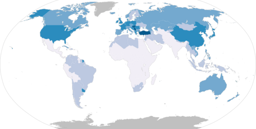
肺がんは、世界的に最も診断数が多く、かつ死亡率の高いがんの一つです。2020年の統計では、世界で220万人が新たに診断され、180万人がこの病気で命を落としています。かつては稀な疾患でしたが、20世紀に入り喫煙が普及したことで急増しました。現在は、早期発見と個別化された治療法の進歩により、生存率の向上が期待されています。
Key Facts
- 主要な原因: 喫煙が全体の80%〜90%を占める最大の要因です。
- 発症年齢: 40歳以降に多く見られ、平均診断年齢は70歳です。
- 主な症状: 咳、血痰、呼吸困難、胸痛などが挙げられます。
- 分類: 主に「非小細胞肺がん(NSCLC)」と「小細胞肺がん(SCLC)」に分けられます。
- 生存率: 5年生存率は世界的に10〜20%ですが、日本は約33%と高い傾向にあります。
肺がんの主な原因とリスク要因
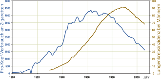
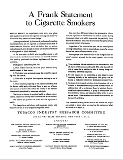
肺がん発症の最大の要因はタバコです。タバコの煙に含まれるニトロサミンなどの発がん性物質(少なくとも50種類が特定されています)がDNAに損傷を与え、細胞をがん化させます。また、本人が吸わない「受動喫煙」であっても、同居家族が喫煙者の場合はリスクが24%上昇するとされており、非喫煙者の肺がんの約17%が環境中のタバコ煙に起因しています。
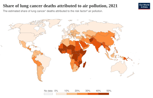
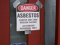
喫煙以外にも、環境的な要因が深く関わっています。大気汚染や、建築資材などに含まれるアスベスト(石綿)、土壌や岩石から自然に発生するラドンガスへの曝露がリスクを高めます。
種類と診断プロセス
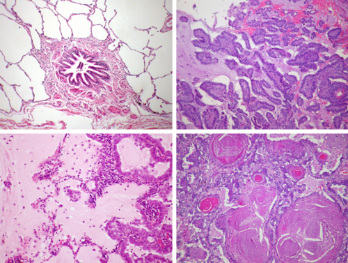
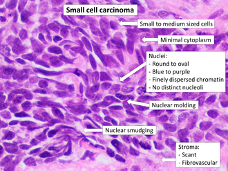
肺がんは、顕微鏡で見た細胞の形態によって大きく2つのタイプに分類されます。一つは進行が非常に速く攻撃的な小細胞肺がん(SCLC)、もう一つはより一般的で多様な形態を持つ非小細胞肺がん(NSCLC)です。NSCLCには、さらに腺がんや扁平上皮がんなどが含まれます。
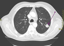
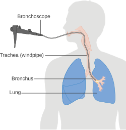
診断には、CTスキャンなどの画像診断と、組織の一部を採取して調べる生検(バイオプシー)が用いられます。生検では、免疫組織化学染色という手法を使い、特定のタンパク質(マーカー)を検出することで、がんの正確な種類を特定します。
ステージ判定(TNM分類)
治療方針を決定するために、TNM分類という世界共通の基準で進行度(ステージ)を判定します。これは、原発腫瘍の大きさや浸潤度(T)、リンパ節への転移状況(N)、遠隔転移の有無(M)を組み合わせて評価するものです。
| ステージ | 特徴(簡略) | 5年生存率 (%) |
|---|---|---|
| IA1 | 腫瘍が非常に小さく、転移なし | 92 |
| IB | 腫瘍がやや大きいが、転移なし | 68 |
| IIA〜IIB | 近接リンパ節への転移あり | 60 〜 53 |
| IIIA〜IIIC | 局所的に進行した状態 | 36 〜 13 |
| IVA〜IVB | 他の臓器へ遠隔転移あり | 10 〜 0 |
早期発見のためのスクリーニング
高リスク群(長期間の喫煙歴がある55歳〜80歳など)に対しては、低線量CTスキャンによる定期的な検診が推奨されています。これにより、自覚症状が出る前に腫瘍を発見でき、死亡率を最大20%減少させられるというエビデンスがあります。ただし、偽陽性(がんではないのに陽性と判定されること)による不要な精密検査のリスクも伴います。
治療法とアプローチ
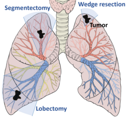
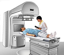
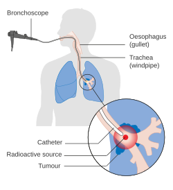
治療はがんの種類とステージに応じて組み合わせて行われます。早期がんの場合は、手術による切除が第一選択となります。また、放射線治療は外部から照射する方法のほか、気道を通じて内部から照射する小線源治療(ブラキセラピー)などの手法があります。
進行がんに対しては、化学療法(抗がん剤)に加え、特定の遺伝子変異を標的とする分子標的薬や、免疫システムを活性化させてがんを攻撃する免疫チェックポイント阻害薬などの最新治療が導入されています。また、身体的・精神的苦痛を緩和する緩和ケアも、診断初期から並行して行われることが重要です。
予後と今後の展望
肺がんの予後は、診断時のステージに強く依存します。早期(IA1)に発見されれば90%以上の高い生存率が見込めますが、末期(IVB)になると極めて困難になります。また、女性は男性よりも早期に発見される傾向があり、生存率が高いことが分かっています。
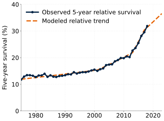
現在、世界中で数千件の臨床試験が進んでおり、特に放射線療法の最適化や、EGFR、VEGFなどの新たな標的分子を用いた薬剤の開発が加速しています。
Frequently Asked Questions
タバコを吸わない人でも肺がんになりますか?
はい、なります。非喫煙者の肺がんの多くは、受動喫煙や大気汚染、アスベスト、ラドンなどの環境要因、あるいは遺伝的な要因によって引き起こされます。
低線量CT検診は誰が受けるべきですか?
一般的に、55歳から80歳の間で、30パックイヤー(1日1箱を30年など)以上の喫煙歴がある高リスク者が推奨されます。詳細な基準は医師にご相談ください。
小細胞肺がんと非小細胞肺がんの違いは何ですか?
小細胞肺がんは進行が非常に速く、転移しやすい傾向がありますが、化学療法や放射線治療への初期反応が良いのが特徴です。非小細胞肺がんは種類が多様で、早期であれば手術による完治の可能性があります。
肺がんを予防するためにできることはありますか?
最も効果的な方法は禁煙することです。また、受動喫煙を避けることや、アスベストなどの有害物質が含まれる環境への曝露を最小限に抑えることが重要です。
ステージIVになっても治療の意味はありますか?
はい。完治が難しい場合でも、分子標的薬や免疫チェックポイント阻害薬などの治療により、がんの進行を遅らせ、生活の質(QOL)を維持しながら生存期間を延ばすことが可能です。
References
- , "Epidemiology".
- , "Age".
- , "Lung cancer".
- , "Introduction".
- , "Presentation/Initial Evaluation".
- , "Clinical Manifestations".
- , "Clinical Manifestations".
- "Diagnosis – Lung Cancer". . 1 November 2022. Retrieved 30 November 2022.
- "Lung Carcinoma: Tumors of the Lungs" (online ed.). Merck Manual Professional. July 2020. Retrieved 21 July 2021.
- , "Noninvasive Staging".
📸 フォトギャラリー